Author name: annm1nty

Proximal Femur: Bloodflow and Fractures

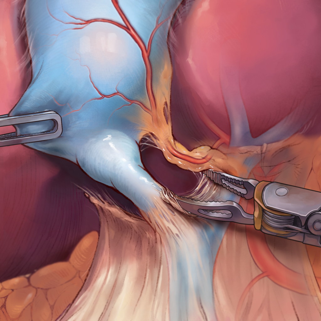
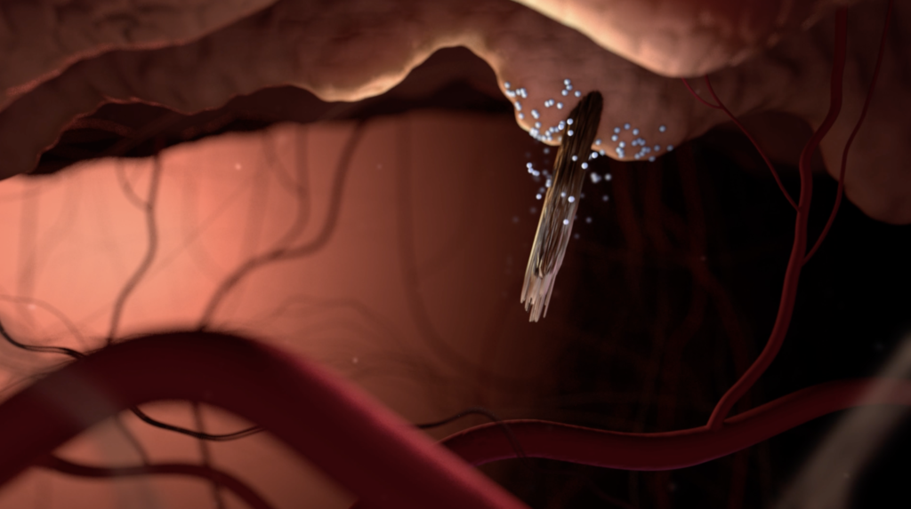
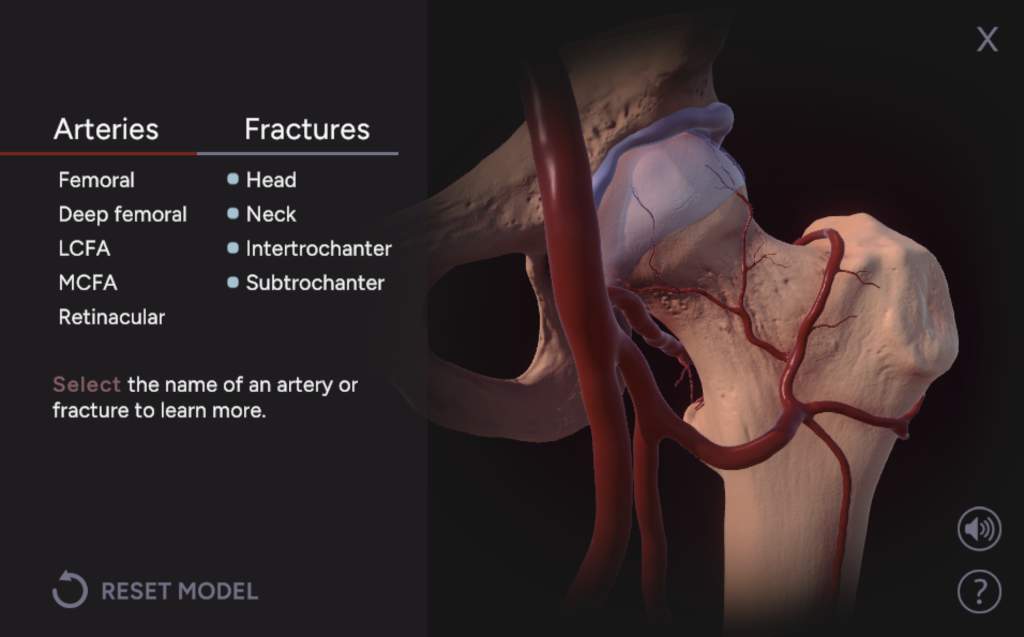
Proximal Femur: Bloodflow and Fractures CLIENT Jeff Day, Jennifer Fairman YEAR 2023 MEDIA Zbrush, Adobe Surface Painter, Unity DESCRIPTION The outcome for patients who su er hip fractures can be dependent on how the fracture impacts bloodflow to the proximal femur. This interactive describes four types of hip fractures and the arteries supplying the proximal femur. An introductory page provides brief context for the interactive module. The main interactive allows users to rotate, zoom in, and click on a three-dimensional model. A side bar lists the names of fracture types and arteries. When a name is clicked, it is highlighted in the model and a brief description appears in the side bar. By viewing selected arterial and bony structures alongside the text description, users can correlate anatomy with potential fracture outcomes.

Proximal Femur: Bloodflow and Fractures Read More »

Husbandry Considerations for Choloepus Senses

Husbandry Considerations for Choloepus Senses CLIENT National Aquarium, Jeff Day, Tim Phelps YEAR 2023 MEDIA Procreate, Adobe Photoshop DESCRIPTION Two-toed sloths (Choloepus) have limited vision and hearing, which informs the way human caretakers interact with them. Created in collaboration with the National Aquarium, this poster describes Choloepus vision and hearing for an audience of people involved in sloth husbandry and an informed lay audience. The top illustration shows the rarely-illustrated eye anatomy of a sloth, depicted in the orbit. An inset compares models of full color vision and Choloepus vision. Two sloth portraits contextualize the orbit and external ear. The bottom banner describes best practices for sloth caretakers.

Husbandry Considerations for Choloepus Senses Read More »

Deep Structures of the Telencephalon

Deep Structures of the Telencephalon CLIENT Lydia Gregg, Mary Ann Wilson YEAR 2023 MEDIA Graphite, Adobe Photoshop DESCRIPTION This illustration was developed as part of a dissection guide to clarify the position of deep brain structures, which are partially visible at the end of the dissection. First, an L-shaped inset provides a step-by-step guide to access the deep structures of the diencephalon. To depict the position of each cut, three-dimensional colored boxes highlight the cortex and white matter removed in each step. External anatomy and cortical regions are highlighted as they are encountered in dissection. The main illustration depicts the anatomic relationships between the deep periventricular structures of the telencephalon in the final stage of the dissection. The overlying cortex, caudate nucleus, thalamus, and amygdala were rendered as partially transparent to clarify the position of these structures and show the arrangement of the hippocampus and fornix. Didactic color was used to help students visualize the position of these structures.

Deep Structures of the Telencephalon Read More »

Cholecystectomy

Robot-assisted Cholecystectomy CLIENT Corinne Sandone, Jennifer Fairman YEAR 2023 MEDIA Adobe Photoshop, Procreate DESCRIPTION This illustration depicts a key surgical moment from a robot-assisted cholecystectomy. In this procedure, the cystic duct and cystic artery are clipped and divided in order to remove the gallbladder. Figure 4a shows the critical view of safety, where the gallbladder has been partially mobilized and the surface of the liver is visible between the cystic duct and artery. Observation of the critical view of safety prevents accidental clipping of the surrounding biliary tree and arteries, which are ghosted in. Figure 4b shows the subsequent division of the cystic duct and artery. This piece was designed for slide format. Surgical Sketches I created sketches of the entire procedure based on my experience observing the surgery in the operating room.

Cholecystectomy Read More »

The Inflammatory Response

The Inflammatory Response CLIENT David Rini YEAR 2023 MEDIA Cinema 4D, Adobe Photoshop, Adobe After Effects DESCRIPTION This animation depicts the physiological and molecular events that occur during an acute inflammatory response through a sequential narrative. It illustrates the key cellular components of inflammation and symptoms that accompany it. By linking these mechanisms to recognizable physiological reactions, the animation provides an overview of inflammation at an appropriate level for the target audience. The narrative uses a pixel game-inspired scene as a framing device to introduce the narrative to viewers with playful, recognizable imagery. The molecular environment is stylized as a simplified, aqueous environment to reduce cognitive load. Storyboards Ahead of creating the animation, I created storyboard sketches in Procreate to establish the content and flow of the video. Process I created my 3D scenes in Cinema4D, while I created frames for the 2D scenes in Photoshop to mimic the look of a pixel game. The final video was created in After Effects, where I put together the animations, voice over, and sound effects.

The Inflammatory Response Read More »

Master’s Thesis: Why Shed the Shield?

Why Shed the Shield? Preceptor Dr. M. Mahesh Faculty Advisor Lydia Gregg YEAR 2024 MEDIA Adobe Illustrator, Photoshop, After Effects DESCRIPTION Gonadal shielding, or placing a protective barrier between patients and a source of radiation to reduce exposure to the gonads, has long been a standard practice in diagnostic imaging. However, advances in technology and new evidence regarding radiation exposure have led the American Association of Physicists in Medicine (AAPM) and other organizations to recommend discontinuation of gonadal shielding. In response, medical facilities nationwide have begun to adopt “no-shield policies,” including the Johns Hopkins Hospital in 2023. The burden of implementing this significant policy shift falls on radiology technologists (RT), who must mediate conversations with patients and caregivers during exams. This thesis project investigated the use of video narrative to convey the scientific rationale behind no-shield policies in an educational campaign for RTs. Storyboards Storyboards were created in Procreate to define the visual content, movement, and overall flow of the animation. Storyboards were revised based on preceptor, advisor, and test audience feedback. Full storyboard available at Jscholarship upon thesis publication. Style & Asset Creation Artwork was created in Adobe Illustrator and Photoshop. Assets for the animation include models for the technologist characters, background art of the hospital setting, and vector illustrations for the scientific content.  Character reference sheet for a technologist. Character reference sheet for a technologist. Vector asset prepared for After Effects animation. Character model prepared for rigging in After Effects.

Master’s Thesis: Why Shed the Shield? Read More »

Engineering Immunocytokines

Engineering immunocytokines to promote anti-tumor activity CLIENT Fabian de Kok-Mercado, Lydia Gregg, Jamie Spangler YEAR 2023 MEDIA PyMOL, Cinema 4D, Adobe Photoshop DESCRIPTION The engineered F10 immunocytokine is composed of an F10 antibody linked to interleukin-2 (IL-2). The structure of the immunocytokine favors the binding of IL-2 to the IL-2 receptor heterodimer on e ector T cells, which triggers a pathway that promotes antitumor activity. This illustration depicts this interaction in an immersive tumor cell environment. The illustration was created for a journal cover, and molecule colors were chosen to align with the accompanying paper’s figure legend. The anatomy of the immunocytokine is labeled, as well as the beta and gamma subunits of the IL-2 receptor. The secondary structure of the subunits, a highlight of the accompanying paper, are included in the illustration. An inset shows a zoomed-out view of an e ector T cell on a tumor cell to contextualize the main image.

Engineering Immunocytokines Read More »

The Optic Nerve Head

The Optic Nerve Head CLIENT Lydia Gregg, Ian Pitha YEAR 2023 MEDIA Graphite, Adobe Photoshop DESCRIPTION This illustration depicts the anatomy of the optic nerve head as it pertains to normal function and glaucoma. First, the main image’s view and orientation are introduced by the inset in the top left. In the main image, a wedge-shaped section of the eye has been removed to show the anatomic relationships between the optic nerve axons, lamina cribrosa, and peripapillary sclera. The section view is offset to reveal the path of the central retinal vessels. To the right of the main image, insets describe the pathology of glaucoma. The top right inset depicts excavation of the optic nerve head. Graphic cellular depictions at the bottom right show the distribution of fibroblasts and glial cells within the nerve axons and extracellular tissue before and after glaucoma, highlighting the extensive cell death seen with excavation.

The Optic Nerve Head Read More »

Scroll to Top