
Deep Structures of the Telencephalon
CLIENT
Lydia Gregg,
Mary Ann Wilson
YEAR
2023
MEDIA
Graphite, Adobe Photoshop
DESCRIPTION
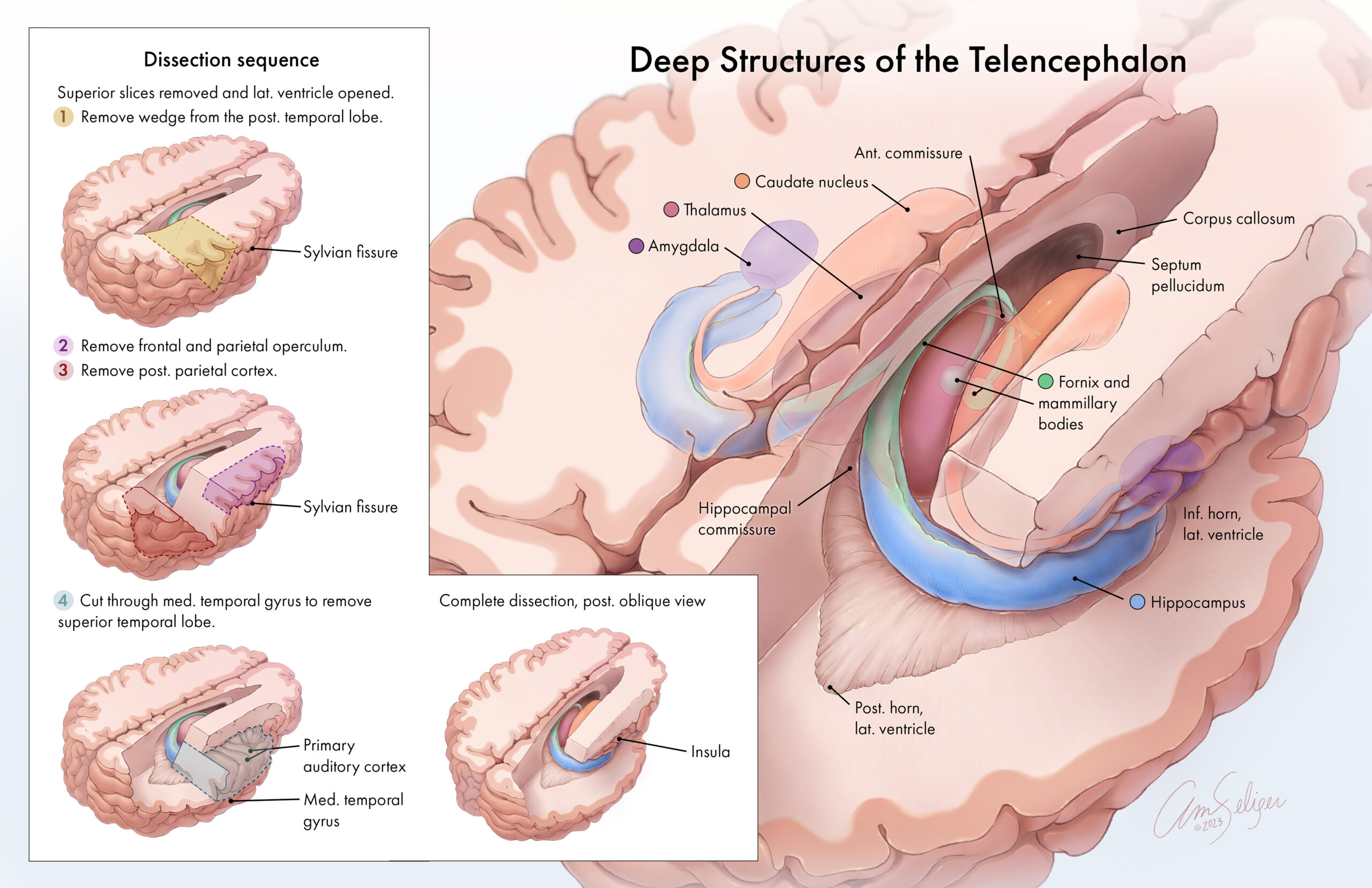
This illustration was developed as part of a dissection guide to clarify the position of deep brain structures, which are partially visible at the end of the dissection. First, an L-shaped inset provides a step-by-step guide to access the deep structures of the diencephalon. To depict the position of each cut, three-dimensional colored boxes highlight the cortex and white matter removed in each step. External anatomy and cortical regions are highlighted as they are encountered in dissection. The main illustration depicts the anatomic relationships between the deep periventricular structures of the telencephalon in the final stage of the dissection. The overlying cortex, caudate nucleus, thalamus, and amygdala were rendered as partially transparent to clarify the position of these structures and show the arrangement of the hippocampus and fornix. Didactic color was used to help students visualize the position of these structures.