
The Optic Nerve Head
CLIENT
Lydia Gregg, Ian Pitha
YEAR
2023
MEDIA
Graphite, Adobe Photoshop
DESCRIPTION
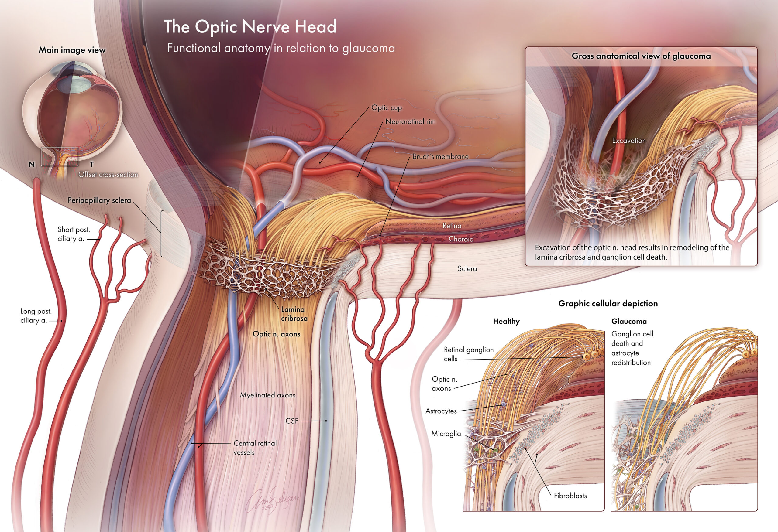
This illustration depicts the anatomy of the optic nerve head as it pertains to normal function and glaucoma. First, the main image’s view and orientation are introduced by the inset in the top left. In the main image, a wedge-shaped section of the eye has been removed to show the anatomic relationships between the optic nerve axons, lamina cribrosa, and peripapillary sclera. The section view is offset to reveal the path of the central retinal vessels. To the right of the main image, insets describe the pathology of glaucoma. The top right inset depicts excavation of the optic nerve head. Graphic cellular depictions at the bottom right show the distribution of fibroblasts and glial cells within the nerve axons and extracellular tissue before and after glaucoma, highlighting the extensive cell death seen with excavation.